Advanced Web-Based DICOM Viewer for Clinical Research
Access, visualize and quantify medical images from any browser with our high-performance zero-footprint and full-featured DICOM viewer.
Professional Imaging Visualization: The Pixilib DICOM Viewer
"In clinical trials and real-world evidence studies, the ability to access imaging data quickly and securely is paramount."
Pixilib's DICOM viewer is a professional-grade, web-based solution designed to provide the capabilities of a high-end workstation directly within your browser.
As a zero-footprint DICOM viewer, it requires no local installation, ensuring seamless access for investigator sites, monitors, and central readers while maintaining the highest standards of data security and regulatory compliance.
From multi-modal DICOM rendering to integrated quantification and AI-assisted segmentation, the Pixilib viewer is the unified visualization layer that powers all our research platforms — letting researchers, radiologists and central readers focus on science, not on software.
Core Features for Expert Medical Review
Built for clinical-grade analysis: the speed of a local workstation, the convenience of a web app, and the precision required for research-grade quantification.
Massive rendering performances
Pixilib's viewer is based on the Open Health Imaging Foundation framework, for which Pixilib is a major contributor. It belongs to a new generation of DICOM web-based viewers — its major innovation lies in its ability to use local hardware resources (including GPU acceleration) to generate every visualization client-side.
While previous generations of web DICOM viewers were slow because of latency to retrieve each visualization from the server, our viewer is extremely smooth — even smoother than professional clinical-grade workstations.
At Pixilib that's our philosophy: no compromise. Research needs the best of image processing to unlock all the capabilities of medical imaging and build a new era of innovative uses. Many research platforms integrate "basic" viewers for basic and generic quantifications — we want to make the most of medical imaging.
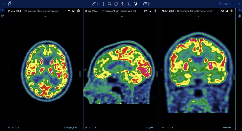
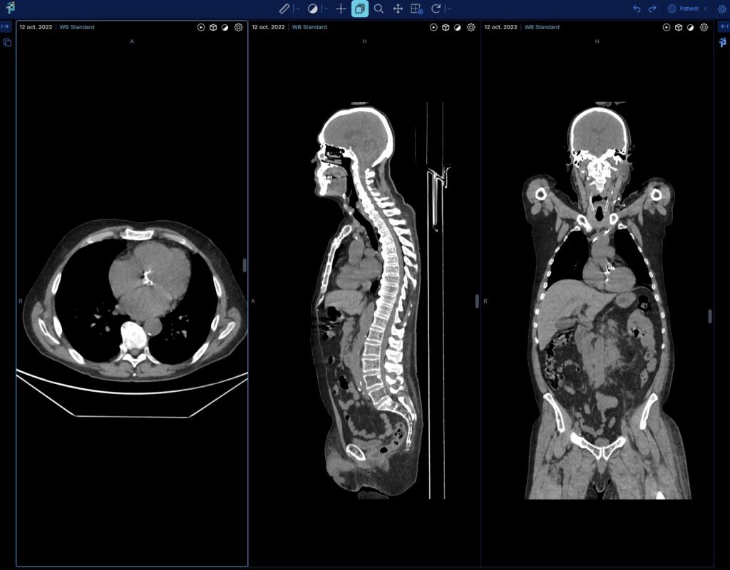
Multi-Modal DICOM Viewer
Our DICOM viewer supports the full spectrum of medical imaging modalities used in modern research:
- Radiology CT, MRI, Ultrasound and X-ray with advanced MPR (Multi-Planar Reconstruction) and MIP (Maximum Intensity Projection)
- Nuclear Medicine seamless PET/CT and PET/MRI fusion with dynamic image support and customizable colormaps
- Dynamic imaging 4D CT, MRI and PET/CT supported with cinematic visualization
- Specialized data Video (Endoscopy), Digital Microscopy (WSI) and Radiotherapy objects (RTSTRUCT, RTDOSE)
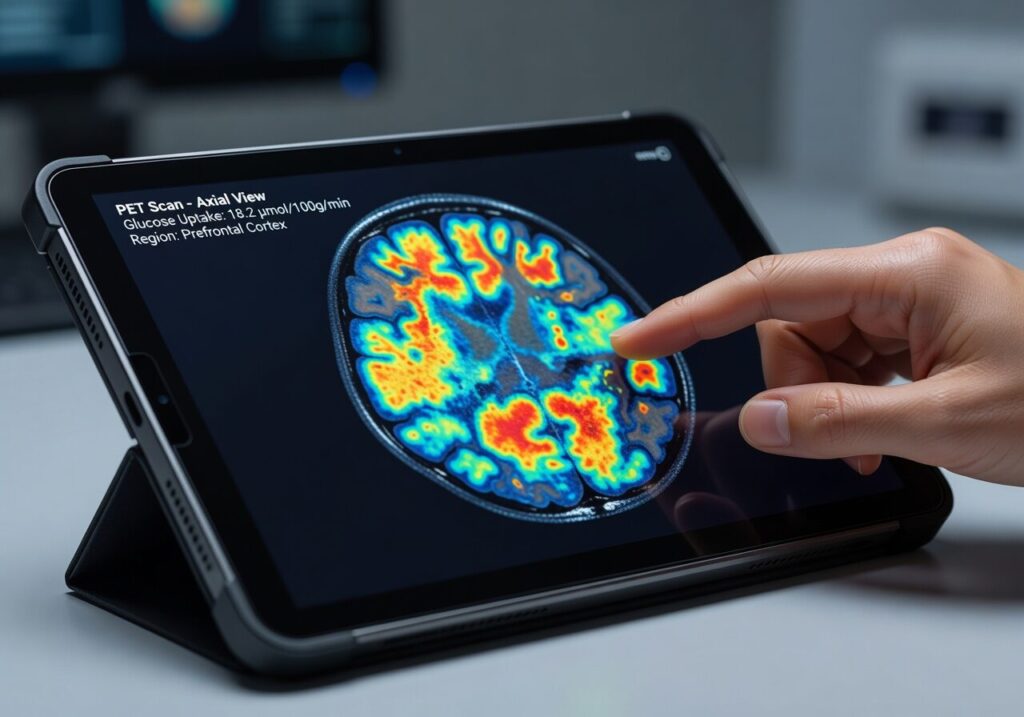
Integrated Quantification & Segmentation Tools
Beyond simple visualization, the Pixilib DICOM viewer is a powerful analytical tool. It integrates advanced quantification features such as:
- Standardized measurements SUV (Standardized Uptake Value) for PET, Hounsfield Units (HU) for CT, distance and angle measurements
- AI-assisted segmentation integration of our AI algorithms for Total Metabolic Tumor Volume (TMTV) or organ segmentation directly within the viewer interface
- Customizable eCRFs perform central review and fill out study-specific forms simultaneously in a unified workspace across GaelO and GaelO-Flow
Native integration in our products
The Pixilib viewer isn't a separate tool you launch on the side — it is the visualization core embedded across the entire Pixilib ecosystem, giving every user the same powerful workspace.
GaelO
In GaelO, our viewer is natively integrated. When opening medical imaging for centralized interpretation, the context is passed from GaelO to the viewer and the physician can fill the personalized eCRF defined for the clinical trial.
Measurements and quantifications flow from the viewer to the eCRF — removing the need for manual copy of data, eliminating typing or rounding errors. Every reviewer uses the same viewer, ensuring standardization of workflow and feature availability for all users, anytime, anywhere.
GaelO-Flow
GaelO-Flow also integrates natively with our viewer. As with GaelO, we integrated customizable CRFs in GaelO-Flow to benefit from standardized data collection in retrospective studies — just like prospective clinical trials.
Researchers can navigate, segment and quantify directly within the same browser session that orchestrates DICOM data flows.
PixiLearn
Pixilib is also involved in education with its PixiLearn platform. The viewer is integrated to deliver immersive teaching with hands-on clinical cases.
We built a remote control system allowing the teacher to highlight areas of interest in the image to participants — a great example of our development capabilities: we start from the use case and build the solution around it.
Why Choose a Zero-Footprint DICOM Viewer?
No installation, no IT bottleneck, no device lock-in — the freedom to focus on imaging research instead of software logistics.
Access without prior Installation
Because it is a zero-footprint DICOM viewer, Pixilib's viewer eliminates the technical barriers often found in multicenter studies. No IT department intervention, no software installation at the recruiting centers — a simple login provides instant access to clinical-grade imaging tools.
Portability
You can start to work on a device and continue later on another one. Our platforms and our viewer are accessible wherever you are — as long as you have a computer with internet access, our viewer will follow you and give maximum freedom to organize your research projects.
Multiple Device Support
Pixilib's viewer can be used on various devices — desktop or laptop computers, tablets or smartphones. Choose your device, our viewer will follow your hardware choice.
A unified infrastructure for medical imaging research
From clinical trial centralization to research PACS and AI segmentation — three complementary products covering the full lifecycle of medical imaging in clinical research.
GaelO
Collection and centralized reading of clinical trials. From DICOM upload to Blind Independent Central Review with disease-specific criteria — GDPR/HIPAA compliant, fully auditable.
Discover GaelOGaelO Flow
Research PACS for retrospective and Real World Evidence studies. Orchestrate medical imaging data: query/retrieve, batch de-identification, AI inference pipelines and seamless export.
Discover GaelO Flow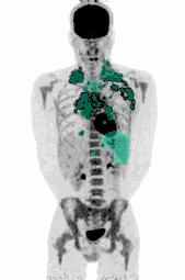
AI Segmentation Algorithms
FDG, PSMA, DOTATOC. Automated PET segmentation to extract Total Metabolic Tumor Volume (TMTV) human-in-the-loop, 100% medically validated, regulatory-ready.
Explore AI algorithms