Advanced Radiomics & Imaging Biomarkers Calculation Services
Extract deep quantitative data from clinical images using state-of-the-art image processing pipelines.
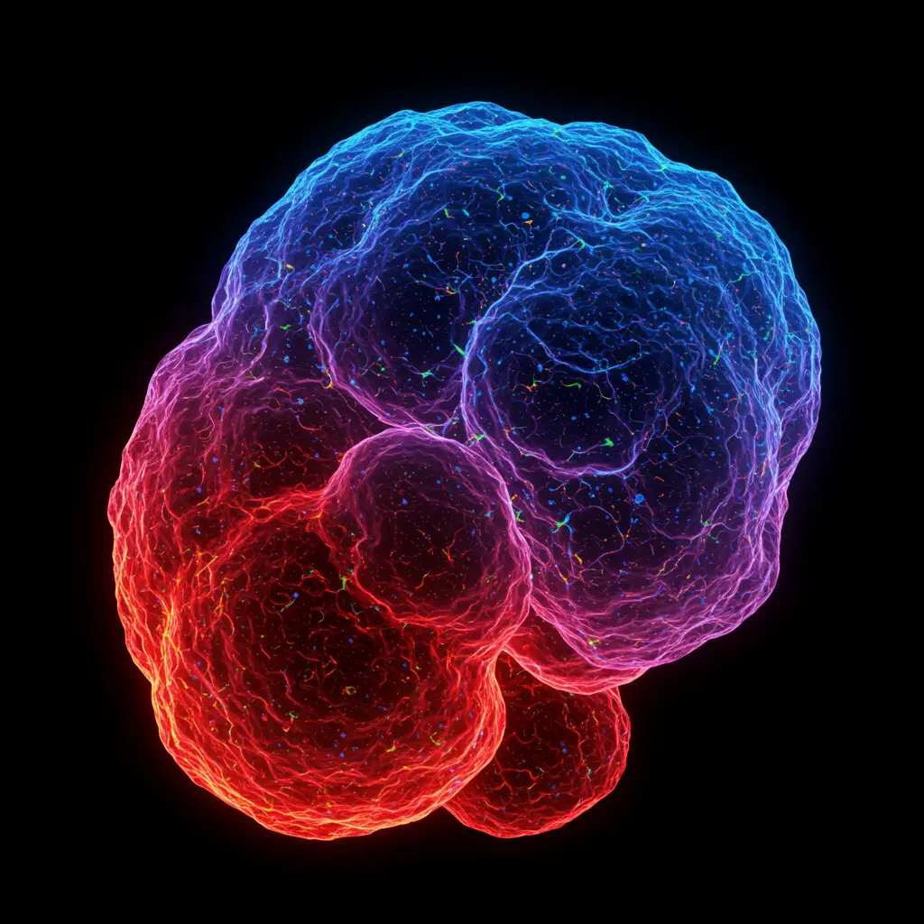
Beyond Qualitative interpretation: The Era of Quantitative Imaging
"We compute texture, shape, and intensity distributions to create new imaging biomarkers that correlate with patient outcomes."
In modern oncology and neurology trials, traditional measurements (like RECIST or PERCIST) are accurate but capture only a fraction of medical imaging capabilities.
We provide comprehensive Radiomics services to identify sub-visual imaging biomarkers and create new biomarkers able to predict the patient's prognosis and evaluate treatment response.
We also apply advanced image processing to medical imaging: we compute texture, shape, and intensity distributions to create and validate new imaging biomarkers that correlate with patient outcomes and could drive better patient care for tomorrow.
Our Radiomics & Image Processing Services
We provide a full-service pipeline to ensure your imaging biomarkers are reproducible, standardized and clinically relevant.
Clinically relevant Radiomics
At Pixilib we know how it can be easy to generate numbers from images and... how it can be clinically irrelevant!
At Pixilib we believe that extracted features should have a biological meaning. We aim to use medical imaging to understand biological mechanisms — we select imaging features that tell disease characteristics or patient condition, not just meaningless numbers.
Radiomics calculations services for prospective and retrospective imaging data
We are ready to analyze medical imaging datasets for various project holders:
- Industrialsanalysis of existing datasets, either prospective or retrospective
- International collaboration projects
- Academic projects
In your datasets we are ready to perform:
- Medical expertisespecific to your research project
- Image segmentationwith our AI tools
- Radiomics calculationsand custom quantifications
We can include our service also with our tools — for instance calculation of radiomics in real time during a trial using the GaelO platform.
High-Fidelity Image Processing & Pre-processing
Before extraction, we take care of data harmonization. Our image processing routines include:
- Resampling & Normalizationensuring consistency across different scanner manufacturers (SUV standardization...)
- Denoising & Filteringdepending on image inputs
- Automated and visual Quality Controlwe check data quality to ensure unbiased results
Radiomics Feature Extraction & Qualification
Using our proprietary image processing engine, we extract hundreds of quantitative features:
- First-order Statisticshistogram-based intensity distribution (Skewness, Kurtosis)
- Morphology / Geometry Quantificationsshape-based descriptors (Sphericity, Surface-to-Volume ratio...)
- Texture Analysis (Gradients)spatial relationship between pixels (GLCM, GLSZM, NGTDM) to characterize tumor heterogeneity
- Custom radiomics depending on project needsdepending on your research project we can evaluate and propose new quantification approaches
Validating Imaging Biomarkers for Clinical Endpoints
The ultimate goal of our Radiomics service is to provide actionable insights. We assist research teams in:
Feature Selection
Identifying the most stable, informative and clinically relevant features to avoid overfitting.
Signature Development
Building multi-parametric models that act as predictive or prognostic imaging biomarkers.
Correlation Studies
Linking radiomic signatures with other patient data (biology, pathology, clinical context...) or clinical survival data.
Why Outsource Your Image Processing to Pixilib?
Three reasons sponsors and academic teams partner with us for their radiomics needs.
IBSI Compliance
Our extraction pipelines follow the Image Biomarker Standardisation Initiative (IBSI) guidelines.
Multimodal Expertise
Radiomics services available for all kinds of imaging modalities — in particular CT, MRI and PET imaging.
Scientific Support
We don't just deliver a CSV file — we help you build and interpret the results for your clinical research.
A unified infrastructure for medical imaging research
From clinical trial centralization to research PACS and AI segmentation — three complementary products covering the full lifecycle of medical imaging in clinical research.
GaelO
Collection and centralized reading of clinical trials. From DICOM upload to Blind Independent Central Review with disease-specific criteria — GDPR/HIPAA compliant, fully auditable.
Discover GaelO
GaelO Flow
Research PACS for retrospective and Real World Evidence studies. Orchestrate medical imaging data: query/retrieve, batch de-identification, AI inference pipelines and seamless export.
Discover GaelO Flow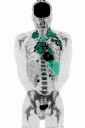
AI Segmentation Algorithms
FDG, PSMA, DOTATOC. Automated PET segmentation to extract Total Metabolic Tumor Volume (TMTV) human-in-the-loop, 100% medically validated, regulatory-ready.
Explore algorithms