Advanced CT Organ Segmentation & Body Composition Analysis
Automate precise anatomical contouring to standardize endpoints and body composition metrics in clinical research.
Transforming Structural Imaging through Automated Organ Segmentation
"In modern clinical trials, structural CT imaging is the backbone of patient monitoring."
Pixilib provides high-performance algorithms for Organ segmentation, enabling rapid, reproducible and clinical-grade anatomical analysis. By leveraging Artificial Intelligence Medical Imaging, our platform detects and contours organs and tissues.
This algorithms allows innovative projects such as:
- Differentiation of local, locoregional and distant involvement of tumors
- Body composition studies (lean tissue, fat tissues perivisceral or subcutaneous, visceral mass...)
- Automated quantification of uptake backgrounds on PET/CT (mediastinum, liver...)
- Organ auto-labelling of PET/CT uptakes
Comprehensive Body Composition & Biomarker Extraction
From sarcopenia studies to opportunistic bone-health screening and radiotherapy planning — one CT segmentation pipeline, multiple research-grade endpoints.
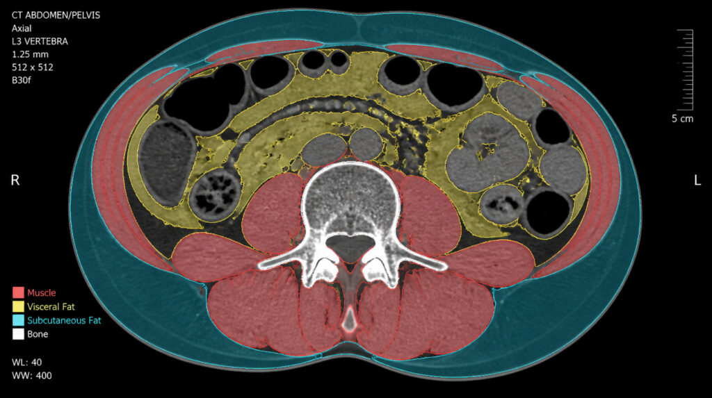
Quantitative Body Composition Metrics
Pixilib's CT segmentation algorithm enables automated Body composition analysis. Our algorithms accurately segment:
- Skeletal Muscle Mass essential for studying sarcopenia and its impact on therapy tolerance
- Adipose Tissue (Visceral & Subcutaneous) — providing critical insights into metabolic health and systemic inflammation
- Bone Density automated vertebral segmentation for opportunistic screening of bone health
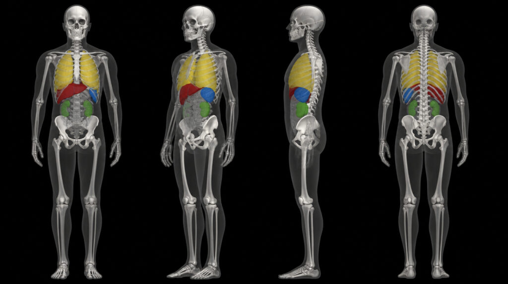
Precise Organ Segmentation for Dosimetry & Safety
For radiotherapy and theranostics, protecting organs at risk (OARs) is vital. Our Organ segmentation engine provides automated contouring of critical structures (liver, kidneys, spleen, lungs), facilitating accurate dosimetry and safety assessment throughout the clinical trial.
Why Choose GaelO for CT Image Processing?
Built around real research workflows: expert-validated AI and zero-friction integration with the platforms you already use.
Expert Validation: Human in the Loop
At Pixilib, we don't believe in fully autonomous "black box" systems for medical research. Every Organ segmentation and Body composition map generated by our AI is presented to the radiologist for review. The physician remains in control, with the ability to adjust contours to ensure 100% accuracy for your scientific publications.
Seamless Workflow Integration
Our CT algorithms are fully integrated into our research platforms, GaelO and GaelO Flow. Once your DICOM data is in our platform, the segmentation pipeline triggers automatically, producing ready-to-use NIfTI or DICOM SEG files for your statistical analysis.
A unified infrastructure for medical imaging research
From clinical trial centralization to research PACS and AI segmentation — three complementary products covering the full lifecycle of medical imaging in clinical research.
GaelO
Collection and centralized reading of clinical trials. From DICOM upload to Blind Independent Central Review with disease-specific criteria — GDPR/HIPAA compliant, fully auditable.
Discover GaelOGaelO Flow
Research PACS for retrospective and Real World Evidence studies. Orchestrate medical imaging data: query/retrieve, batch de-identification, AI inference pipelines and seamless export.
Discover GaelO Flow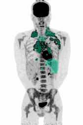
AI Segmentation Algorithms
FDG, PSMA, DOTATOC. Automated PET segmentation to extract Total Metabolic Tumor Volume (TMTV) human-in-the-loop, 100% medically validated, regulatory-ready.
Explore algorithms