Precision Algorithms for Medical Imaging Segmentation
Integrate new imaging biomarkers in research projects and clinical trials with our Artificial Intelligence Medical Imaging processing tools.
Transforming DICOM Data through Advanced Medical Imaging Segmentation
"At Pixilib, we believe that the value of clinical data lies in qualitative evaluation but also in its quantification."
Traditional manual contouring is time-consuming and subject to high inter-reader variability. Our suite of algorithms for medical imaging segmentation is designed to provide accessible, reproducible, high-fidelity results for multicenter clinical trials and real-world evidence studies.
By integrating Artificial Intelligence Medical Imaging into your workflow, we empower researchers to move beyond qualitative assessment. Our AI engines detect, contour, and quantify lesions in seconds, providing a robust foundation for imaging biomarkers and radiomics extraction.
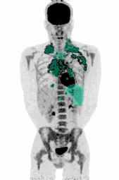
AI-Driven PET Segmentation (Nuclear Medicine)
Nuclear medicine is a relatively confidential area where expertise is rare, especially on topics such as tumor segmentation. We are Nuclear Medicine image processing experts — we built biomarkers on top of PET/CT segmentations. Our medical imaging segmentation tools are optimized for a wide range of radiopharmaceuticals, ensuring consistency across different camera manufacturers and reconstruction protocols.
FDG PET: Automated TMTV Calculation
Automate the extraction of the Total Metabolic Tumor Volume (TMTV). Our algorithms distinguish between pathological uptake and physiological background to facilitate usage of TMTV in clinical trials.
This prognostic biomarker is now integrated in several lymphoma clinical trial imaging protocols to better tailor therapy for patients and is already used as a decisional endpoint. In so many other tumors TMTV is an evolving research topic for which we also aim to create a path to reach clinical practice.
PSMA & DOTATOC: Targeted Tumor Burden Assessment
Precision tools for prostate cancer and neuroendocrine tumors. We leverage Artificial Intelligence Medical Imaging to accurately segment specific tracer uptake, facilitating dosimetry and therapy response monitoring in theranostics.
CT Segmentation: Organ segmentation and Body Composition Analysis
Structural imaging remains the backbone of oncology and radiology research. Pixilib provides dedicated engines for structural medical imaging segmentation.
Automated Organ segmentation
Our AI models provide automated segmentation of organs on CT scans. This technology can be used for several research use cases: differentiate and quantify tumor involvement per organ, or compute body composition calculations.
Body Composition calculation
With our CT segmentation algorithm, we can compute the body composition automatically. We differentiate and quantify:
- Muscular mass (muscle body mass)
- Lean Body mass
- Perivisceral adipose mass
- Subcutaneous adipose mass
- Bone tissue with bone piece-by-piece identification
Anthropometric parameters calculation are readily available in our research platforms, making them widely available for research topics.
Why Choose Pixilib Algorithms for Your Research?
AI built around real research problems, with researchers and medical experts kept firmly in the loop — not "black box" promises, but tools that actually accelerate science.
Trained to solve real problems
At Pixilib we don't train models with promises of a new AI era that will save patients' lives by itself. We train models to help researchers achieve their goal — improving meaningful data extraction from medical imaging to build and validate clinically relevant imaging biomarkers.
With these new biomarkers we aim to create, with researchers, innovative clinical trials that will drive patients' treatment toward new strategies guided by medical imaging. We believe AI is a wonderful and powerful tool as a companion to researchers — keeping human-in-the-loop result validation.
Seamless Integration with GaelO and GaelO Flow platforms
Our segmentation algorithms are fully integrated into our GaelO and GaelO Flow platforms to automate workflows. We customize and execute AI pipelines automatically to generate the automated segmentations that will save hundreds of hours to researchers.
In our tools, researchers don't have to worry about the techniques — we made AI available out of the box, we just ship ready-to-use tools to accelerate research capabilities.
Standardized & Reproducible Biomarkers
Whether for a phase III trial or a large RWE study, our algorithms facilitate image feature extraction — reducing the work burden on researchers and enhancing reproducibility. With our AI tools, researchers can focus on their expertise, validate, make corrections for complicated cases and leverage AI results to the highest level of expertise.
Human in the Loop: The Best of Both Worlds
We don't believe in "black box" AI. Our Artificial Intelligence Medical Imaging tools are designed as an assistant for the researchers. Every segmentation can be reviewed, adjusted, and validated by a medical expert, ensuring a final accurate result for your clinical endpoints.
A unified infrastructure for medical imaging research
From clinical trial centralization to research PACS and AI segmentation — three complementary products covering the full lifecycle of medical imaging in clinical research.
GaelO
Collection and centralized reading of clinical trials. From DICOM upload to Blind Independent Central Review with disease-specific criteria — GDPR/HIPAA compliant, fully auditable.
Discover GaelOGaelO Flow
Research PACS for retrospective and Real World Evidence studies. Orchestrate medical imaging data: query/retrieve, batch de-identification, AI inference pipelines and seamless export.
Discover GaelO FlowAI Segmentation Algorithms
FDG, PSMA, DOTATOC. Automated PET segmentation to extract Total Metabolic Tumor Volume (TMTV) human-in-the-loop, 100% medically validated, regulatory-ready.
Explore algorithms